Butterfly wing magnification series
Voting period is over. Please don't add any new votes. Voting period ends on 30 May 2010 at 14:34:43 (UTC)
Photographic and light microscopic images 

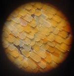
Zoomed-out view of an Aglais io. Closeup of the scales of the same specimen. High magnification of the coloured scales (probably a different species). Electron microscopic images 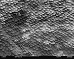


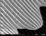
A patch of wing Scales close up A single scale Microstructure of a scale Magnification Approx. ×50 Approx. ×200 ×1000 ×5000


- Original
- The Lepidopteran wing surface is made up of usually coloured scales, shown here at various magnifications. Higher magnifications require scanning electron microscopy to be used, which depicts objects in a greyscale shading.
- Reason
- Illustrates both the structure of a butterfly wing at various scales of magnification, and the relative merits and disadvantages of light vs electron microscopy (for possible later inclusion in microscopy-related articles). All images above current standards. Please review this with fairness towards the contributors rather than the nominator. Thank you.
- Articles in which this image appears
- butterfly, external morphology of Lepidoptera, scale (Lepidoptera)
- FP category for this image
- Wikipedia:Featured pictures/Animals/Insects
- Creators
- Michael Apel (nos. 1 and 2), Shaddack (no. 3), SecretDisc (nos. 4 to 7)
- Support as nominator --Papa Lima Whiskey (talk) 14:34, 21 May 2010 (UTC)
- Support wow, high encyclopedic value. --Alchemist-hp (talk) 14:56, 21 May 2010 (UTC)
- Enthusiastic support Highly interesting, highly encyclopedic, and access to SEMs doesn’t come cheap and Wikipedia would be better off with more of this sort of thing. Greg L (talk) 16:17, 21 May 2010 (UTC)
- Oppose The image nominated below Inachis io Lill-Jansskogen.JPG is much better than image one in this set. Image 3 is very fuzzy, noisy, barely meets the size requirements and is most probably not of the same specie. Images 4-7 all have notable blurry lines. Very good EV but the technicals are weak --Muhammad(talk) 17:13, 21 May 2010 (UTC)
- Strong Support Massive EV in my view; the quality is also excellent for the mediums presented, though a little weaker around image three. I think the third image is, however, forgivable. Cowtowner (talk) 01:14, 22 May 2010 (UTC)
- Comment I love the idea, but I think some color closeups of the scales that are of the same species and better quality would greatly amplify the series... I do admit those may be quite a difficult task to get unless someone here has access to a good universities biology lab and the desire to get the images (And specimens). The SEM set is wonderful, and I do agree with Muhammad that the nominated image below (Inachis io Lill-Jansskogen.JPG) would be a better replacement for the first image in the set. I would change to weak support if the first image was changed, support if that was done and the amateur microscopy image was dropped, and strong support if a better microscopy image could be acquired showing the scales in visible light. — raeky (talk | edits) 18:08, 22 May 2010 (UTC)
- Comment: I'd be inclined to say that just the electron microscope shots would be best- they're quite clearly a "set", while the others are not so much. There are issues with both the first and third shots, and the way you've grouped them in this nomination even shows two distinct subsets. J Milburn (talk) 10:47, 25 May 2010 (UTC)
- Numbers 1 and 2 are FPs on Commons. Papa Lima Whiskey (talk) 14:35, 25 May 2010 (UTC)
- Support: Interesting and High EV. --Redtigerxyz Talk 16:30, 25 May 2010 (UTC)