.mw-parser-output .hidden-begin{box-sizing:border-box;width:100%;padding:5px;border:none;font-size:95%}.mw-parser-output .hidden-title{font-weight:bold;line-height:1.6;text-align:left}.mw-parser-output .hidden-content{text-align:left}@media all and (max-width:500px){.mw-parser-output .hidden-begin{width:auto!important;clear:none!important;float:none!important))You can help expand this article with text translated from
the corresponding article in German. (February 2023) Click [show] for important translation instructions.
View a machine-translated version of the German article.
Machine translation, like
DeepL or
Google Translate, is a useful starting point for translations, but translators must revise errors as necessary and confirm that the translation is accurate, rather than simply copy-pasting machine-translated text into the English Wikipedia.
Consider
adding a topic to this template: there are already 1,897 articles in the
main category, and specifying|topic= will aid in categorization.
Do not translate text that appears unreliable or low-quality. If possible, verify the text with references provided in the foreign-language article.
You must provide
copyright attribution in the
edit summary accompanying your translation by providing an
interlanguage link to the source of your translation. A model attribution edit summary is Content in this edit is translated from the existing German Wikipedia article at [[:de:Heidelberg Retina Tomograph]]; see its history for attribution.
You may also add the template ((Translated|de|Heidelberg Retina Tomograph)) to the
talk page.
For more guidance, see
Wikipedia:Translation.
| Heidelberg Retinal Tomography |
|---|
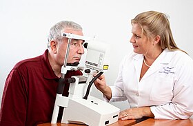 Examination with the Heidelberg Retina Tomograph |
|
The Heidelberg Retinal Tomography is a diagnostic procedure used in ophthalmology. The Heidelberg Retina Tomograph (HRT) is an ophthalmological confocal point scanning laser ophthalmoscope[1] for examining the cornea and certain areas of the retina using different diagnostic modules (HRT retina, HRT cornea, HRT glaucoma). However, the most widely used area of application for HRT is the inspection of the optic nerve head (papilla) for early detection and follow-up of glaucoma. The procedure has established itself as an integral part of routine glaucoma diagnostics alongside the visual field examination (perimetry), the chamber angle examination (gonioscopy) and the measurement of intraocular pressure (tonometry). The HRT is the most widely used application of confocal scanning laser ophthalmoscopy.[2]
Principle
It is based on the principle of spot illumination and spot detection. The optic nerve and retina are illuminated through a single pinhole, and the light returning from the area of interest results in a 2D image.[3] from a series of 2D images, a 3D image is made.[3]
Advantages and disadvantages
Its advantages include rapid image acquisition without the need for pupillary dilation, three-dimensional topographic representation of the optic nerve head, advanced data analysis capabilities built into the machine, and the ability to provide accurate data analysis over time.[3] Also, the technology of HRT is very accurate and test-retest variability is very low.[3] Another important advantage is that it is more comfortable for the patient as the laser only illuminates the area of interest with a low light intensity and short duration (1.6 seconds).[3]
It is necessary to use a reference plane, drawing the contour line depends on the examiner (this is difficult in many eyes where the edge of the disc is not clearly visible), the sensitivity and accuracy decreases in large eyes with myopic changes, and the pulsation of blood vessels may cause deviations in measurements.[3] Changes in intraocular pressure may also affect HRT measurements.[4] These are main disadvantages of HRT.
Glaucoma Diagnosis
During the examination, a laser beam passes through the pupil opening onto the back of the eye and scans the optic nerve head and the retina. A three-dimensional image is generated from several tens of thousands of measuring points, which allows a quantitative assessment of all relevant anatomical structures:[5]
- disc cup (shape, asymmetry),
- neuroretinal rim (area and volume) and
- peripapillary retinal nerve fiber layer (retinal surface height variation, thickness, asymmetry).
These stereometric parameters are compared with extensive databases and thus enable the eye to be classified taking into account the individual papillae size and the patient's age. Two independent classification methods based on different approaches are available.
